ABOUT DEPARTMENT
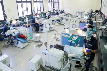
The Department of Conservative dentistry and endodontics is pillared by the ideology of achieving excellence in the field and boasts of the state of the art technology and expertise in meeting this end. Conservative procedures and endodontic procedures performed in the department are in accordance with and the finesse of international standards. The department of Conservative dentistry caters to both undergraduate (UG) and postgraduate (PG) training curriculum. To contribute to the advancement of the field is valued as a high virtue and the department strongly believes in it. The dissertations and the thesis of the postgraduate residents are epitomes of original research and involve areas of international scientific interest including some groundbreaking pioneer studies in the field.
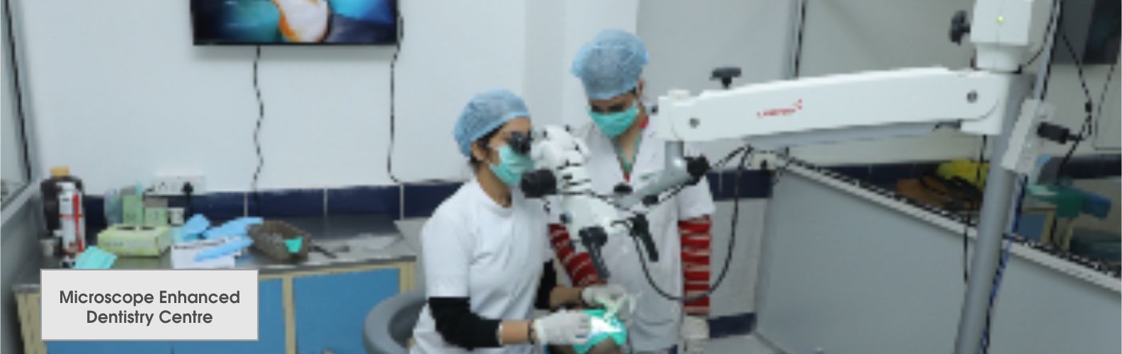
CENTRE FOR MICROSCOPE ENHANCED DENTISTRY
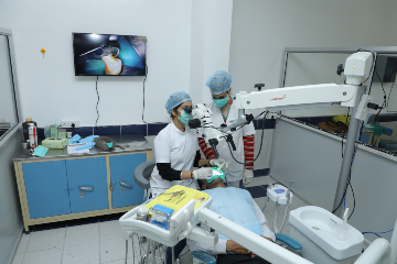
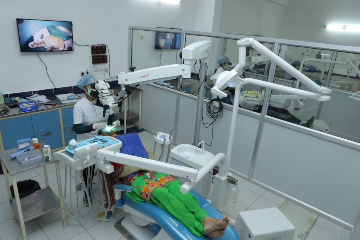
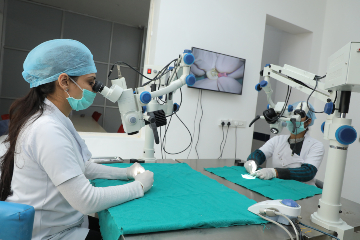
The Centre for Microscope Enhanced Dentistry was established in the Dept. of Conservative Dentistry and Endodontics, FDS, SGT University, Gurugram in January 2019. Dental operating microscope (DOM) is an important tool that allows for better visualization through magnification of dental hard and soft tissues. DOM enhances precision which in turn enhances the quality of work ensuring long-term success.
The Centre for Microscope Enhanced Dentistry is one of its kind in the northern part of the country. It is equipped with multiple clinical dental operating microscopes and preclinical DOM. This Centre was inaugurated by honorable DCI President Dr. Dibyendu Mazumder.
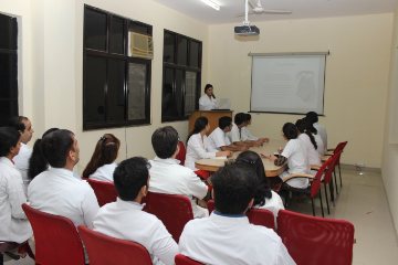
The five basic advantages in using the DOM for an endodontic specialist include: increased visualization, improved quality and precision of treatment, enhanced ergonomics, ease of proper digital documentation and increased communication ability through integrated video. In this Center the postgraduates will learn “How to use Microscope for various clinical procedures.” Basic information regarding handling of microscope is also provided. The preclinical training program is also provided for postgraduates under which they perform their preclinical exercises under microscope. The various procedures done under microscope in our department are composite fillings, inlay and onlay preparations, veneering, tooth build ups, core build ups, endodontic therapy procedures, Re root canal procedures, microsurgical procedures like apicoectomies, hemisection, root amputation, bicuspidization, retro grade fillings, file retrieval procedures, root perforation repairs, apical barrier formation procedures, tooth and crown preparations, detection of cracks, vertical root fractures, detection of hidden canals, identification of MB2 canals, opening of calcified canals, management of endoperio lesions and reattachment of fractured teeth. The enhanced vision with magnification and illumination from a microscope allows endodontist to observe the most coronal aspects of fractured post and broken instruments and to remove them without any major loss of tooth structure and perforations.
With enhanced visualization, the clinician’s ability to diagnose problems in the earlier stages of a disease process is possible. Treatments also can be performed with a greater level of precision, thereby reducing the occurrence of failures and the need for redos. The introduction of the dental operating microscope and the associated ability to inspect the root canals – both orthograde and retrograde – have fundamentally changed our understanding of dental morphology and its complexity. In today’s era of modern endodontics DOM is one of the most important tool in dental armamentarium.