Hemi-Facial Microsomia
Patient reported to the department with complaint of difficulty in eating food due to irregularly arranged teeth.

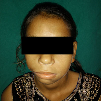
Extra-oral examination revealed prominent forehead, receded hairline, right ear smaller in size (microtia), malformed with a roughly oval shape and 3-4 blind fistulas overlying the ear (atresia). A horizontal scar mark measuring 18-20 mm in size seen extending from the right angle of the mouth till a line drawn perpendicular to the lateral canthus of the eye suggestive of lateral facial cleft. Right side mandible smaller in size, head of the right condyle is not palpable, hypotrophy appreciated in right masseter muscle. Intra oral examination revealed severe malocclusion with interdental spacing between anterior maxillary teeth suggestive of developing malocclusion.

Panoramic radiograph reveals mixed dentition phase with multiple developing tooth buds. Zygomatic process cannot be traced completely on the right side. The right half of the mandible from the symphysis region appears sharply bent upto the angle region. Right side temporo-mandibular joint is not appreciable,

PA skull radiograph revealed an asymmetry on the right side with hypoplastic ramus, hypoplastic condylar head and aplastic coronoid process. The similar findings were confirmed by CT scan.
Final diagnosis: Hemifacial Microsomia on right side OMENS Classification: O0M2BE3N71S2
Management:
Immediate treatment: Orthodontic Opinion
Planned treatment: Mandibular distraction osteogenesis.


