DEPARTMENT OF CONSERVATIVE DENTISTRY AND ENDODONTICS
CLINICAL CASE DISCUSSIONS
One of the most pertinent aspects of operative dentistry and endodontics is the delivering and maintaining a perfect amalgamation of functional continuum with esthetics; and in itself, it requires highest degrees of skill, precision and knowledge.
Overcoming challenges, delivering sound and stable restorations, along with elimination of pathology and root causes, while alleviating pain and crafting esthetically pleasing tooth profiles is our aim; to be delivered to each patient.
The special case report that follows, is a summary of operative and treatment oriented challenges, which were overcome by correct use of diagnostic skills, charting of a conservative line of treatment plan, precise use of skills and knowledge and delivering of a pain free, healthy and sound rehabilitation.
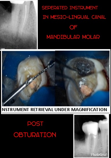
Case 1. RETRIEVAL OF SEPARATED INSTRUMENT FROM THE ROOT CANAL UNDER DOM
Patient presented with severe pain in lower mandibular molar area. Past dental history indicated attempted root canal treatment 2 weeks back. Radiograph revealed instrument separation in distal canal of mandibular molar. With expertise the instrument was retrieved under Dental Operating Microscope. Root canal treatment was completed successfully and the patient bid off smiling with no pain.
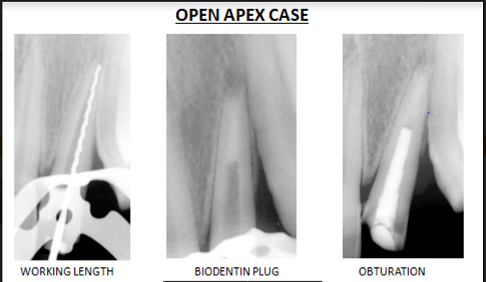
Case 2. MANAGEMENT OF OPEN APEX UNDER DENTAL OPERATING MIOCROSCOPE
The patient came with persistent pain in upper front tooth region post traumatic injury occurred 5 years back. On radiographic examination an open apex was spotted in relation to the upper lateral incisor. The working length was determined with apex locator. Biomechanical preparation was done with rotary file system and the open apex was closed under Dental Operating Microscope with the formation of biodentin plug. Pain was relieved and the patient was kept on follow up.
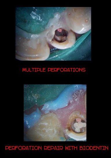
Case 3. PERFORATION REPAIR UNDER DENTAL OPERATING MICROSCOPE
Patient presented with slight numbness and pain on chewing in the lower premolar region due to which he had difficulty in eating food. Radiographic examination revealed attempted root canal treatment. When operated under Dental Operating Microscope multiple perforations were seen. The perforations were sealed with BIODENTIN and the root canal treatment was completed successfully. The patient went back home with proper functioning teeth.
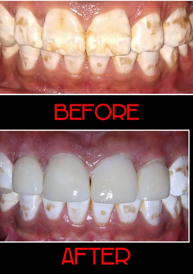
Case 4. Management of discolored teeth with veneers
The patient presented with severe flourosis in front teeth and complained of poor esthetics. Treatment was provided with laboratory fabricated beautiful zirconia veneers with minimal expenditure. The patient gained confidence to smile.

Case 5. Intentional reimplantation
A 23 year old male patient, reported to the department of conservative dentistry and endodontics with chief complaint in lower left back tooth region.
IOPA revealed the unusual anatomy of tooth.- TAURODONT TOOTH.
CBCT report showed that there were three mesial and three distal canals in the tooth and periapical lesion was present.
Root canal therapy was done and intentional REIMPLANTATION procedure performed to seal the apex.

Case 6. Fragment reattachment after traumatic injury
A 22 year old Patient reported with a history of trauma causing fracture of upper anterior teeth few hours back , after thorough examination , a diagnosis of Ellis class III fracture in relation to 11 and 12 was made .Fractured segments were attached palatally at subgingival level . Intensive treatment plan was charted, and after acquiring patient’s consent, reattachment of the fractured segment after root canal treatment was planned on the same day . Fractured segments were extracted atraumatically and placed in normal saline and Single sitting RCT with fibre post was done to relieve the pain. After that gingivectomy was performed in relation to same teeth and fractured segments were reattached to their corresponding position with resin cement . Follow up was done after a month and patient was symptom free and happy.

CASE 7. COMPOSITE VEENERING
The patient reported to the department of Conservative Dentistry & Endodontics with the chief complaint of fractured upper front teeth. The patient was diagnosed with Ellis Class I wrt 11 & Class II fracture wrt 12. The treatment Plan was formulated as direct composite build up. The patient was satisfied with the post operative appearance.

Case 8. Perforating internal resorption
In this case, the patient came with chief complaint of pain in the upper front teeth, on oral examination, the teeth morphology appeared normal, however radiographic examination revealed internal root resorption in relation to 11; further investigations were carried out using the cone beam computed tomography (CBCT) to reveal the extent of the defect and thereby chart the treatment and prognosis of the affected tooth; treatment plan included filling of the defect with glass ionomer cement and sealing the root apex using MTA(Mineral trioxide aggregate), post endodontic therapy.

Case 9. apicoectomy
In this case, the patient presented with pain in the upper front teeth since 10 days and gave a history of trauma 6 yrs back. No history of swelling and medication was given, on intra-oral examination, Ellis class 1 fracture w.r.t 11 & 21 was revealed. Radiographic examination showed a Peri apical radiolucency and an open apex w.r.t 11 & 21.the diagnosis was established as symptomatic peri-apical radiolucency w.r.t 11,21 a treatment plan of peri-apical surgery w.r.t 11 & 21 was charted and carried out.


